
Ptosis Surgery in Turkey — Levator Repair, MMCR and Frontalis Sling at Eyeglow Istanbul
Functional ptosis repair with precise lid measurement before any surgical recommendation. MMCR (no skin incision), levator advancement and frontalis sling — selected by etiology, not by request. Led by our oculoplastic surgical team, with neurogenic and myogenic cases fully evaluated before surgery is offered.
Verified and listed across leading directories and authorities


What is ptosis surgery?
Ptosis surgery (blepharoptosis repair) corrects a drooping upper eyelid caused by levator muscle weakness, aponeurotic disinsertion, or congenital levator dysplasia. Three techniques are used at Eyeglow: MMCR (Müller muscle resection — posterior, no skin incision, for mild ptosis), levator advancement (anterior approach, moderate-to-severe), and frontalis sling (severe cases with absent levator function). The technique is selected after formal levator function measurement and phenylephrine test — never by appearance alone.
Ptosis surgery is distinct from upper blepharoplasty. Blepharoplasty removes excess eyelid skin (dermatochalasis) without changing lid margin height. Ptosis repair elevates a functionally low lid margin by repairing the levator mechanism. When both conditions coexist — excess skin and a true levator deficit — a combined procedure is appropriate. Recommending blepharoplasty for a patient with untreated ptosis will not correct the lid height and is not clinically appropriate. At Eyeglow, every patient is assessed with formal lid measurements (MRD-1, levator function in millimetres, Bell's phenomenon, phenylephrine test) before any surgical option is discussed.
Ptosis surgery is not always elective. Congenital ptosis covering the pupil in a child is an ophthalmic urgency due to amblyopia risk. Sudden-onset ptosis in an adult requires neuro-ophthalmic evaluation before any surgical planning — ptosis from aneurysm or myasthenia gravis requires urgent medical attention, not eyelid surgery. Our surgical team's assessment includes a structured neuro-ophthalmic checklist; surgery is never offered based on photographs alone.
From initial assessment to eyelid symmetry
- 01
Oculoplastic assessment and etiology review
You share upper eyelid photographs from different gaze directions, prior medical records (neurology reports, Tensilon test results if relevant, previous lid measurements) and a description of onset — was the ptosis present from birth, gradual onset in adulthood, or sudden? Our oculoplastic surgical team reviews your case to classify the ptosis type (congenital, aponeurotic, neurogenic, myogenic or mechanical), measure the marginal reflex distance (MRD-1), and determine whether amblyopia risk applies (paediatric cases). A surgical technique is not recommended until the etiology is established — ptosis surgery on an undiagnosed neurogenic or myogenic cause without neuro-ophthalmic clearance is unsafe.
- 02
Pre-operative measurements in Istanbul
On day 1 we perform standardised ptosis measurements: marginal reflex distance (MRD-1 and MRD-2), levator function (LF) in millimetres of lid excursion from downgaze to upgaze, upper lid crease position, Bell's phenomenon (upward globe rotation on lid closure — critical for lagophthalmos risk), corneal sensation, dry eye assessment (Schirmer test, TBUT) and phenylephrine test (to determine Müller muscle responsiveness for MMCR candidacy).
- 03
Technique selection and consent
The surgical technique is matched to levator function and Müller muscle response: MMCR (Müller muscle conjunctival resection) for mild ptosis with good Müller muscle response to phenylephrine; levator advancement or resection for moderate-to-severe ptosis with measurable levator function (≥4 mm); frontalis sling for severe ptosis with absent levator function (LF <4 mm). Combined ptosis repair with upper blepharoplasty is performed when dermatochalasis coexists — the combined approach is explained with its specific indications and not offered as a cosmetic upgrade.
- 04
The procedure (45–90 minutes)
MMCR: performed through a posterior (conjunctival) approach — no visible skin incision. The Müller muscle and conjunctiva are resected en bloc based on the phenylephrine test result; the posterior incision is self-sealing or closed with absorbable sutures. Levator advancement / resection: through the upper lid skin crease incision, the levator aponeurosis is identified, advanced anteriorly and fixed to the tarsal plate with adjustable sutures — lid height is assessed during surgery under local anaesthesia with the patient seated. Frontalis sling: a sling material (silicone rod or autologous fascia lata from the thigh in paediatric cases) is passed through the lid and fixed to the frontalis muscle, allowing the brow to elevate the lid when levator function is absent.
- 05
Day 1, day 5 reviews — asymmetry assessment
Eyelid position is evaluated at day 1 and day 5. Minor asymmetry between the two sides is common in the first week due to swelling; the operated side often appears overcorrected (higher than desired) initially as oedema resolves. If lid height is clearly over- or undercorrected at day 5, early adjustment is possible while tissue is still mobile — levator sutures can be released (overcorrection) or advanced further (undercorrection) within the first one to two weeks.
- 06
Structured follow-up and amblyopia monitoring
Paediatric ptosis patients are followed at one month and three months for amblyopia treatment (patching or penalisation) if visual axis coverage was present. Adult patients are reviewed at one month and six months, with photographs documenting symmetry and lid height. Lagophthalmos (incomplete lid closure) is monitored at every visit — nocturnal lubricant drops are prescribed if residual lagophthalmos exceeds 2 mm. Our surgical team provides written instructions for emergency contact if sudden overcorrection threatens corneal exposure.
MMCR vs Levator Advancement vs Frontalis Sling — how the technique is chosen
The ptosis repair technique is determined by levator function (LF in mm), Beard severity classification and etiology — not by patient preference. Here is how the three approaches differ:
| Aspect | MMCR (Müller Resection) | Levator Advancement / Resection | Frontalis Sling |
|---|---|---|---|
| Technique name | Müller Muscle Conjunctival Resection (MMCR) | Levator Advancement or Resection | Frontalis Sling |
| Approach (incision) | Posterior — through conjunctiva, no visible skin scar | Anterior — through upper lid crease (same as blepharoplasty incision) | Small forehead and lid incisions for sling passage |
| Ideal patient | Mild ptosis (MRD-1 ≥0 mm), good levator function (≥10 mm), positive phenylephrine test (≥1.5 mm elevation with 10% phenyephrine drops) | Moderate to severe ptosis, measurable levator function (4–10 mm LF), aponeurotic or congenital with partial function | Severe ptosis with absent or near-absent levator function (LF <4 mm), including congenital ptosis with amblyopia risk |
| Ptosis correction | 1–3 mm reliable lift; predictable for mild cases with positive Müller response | 2–5 mm lift; adjustable intraoperatively under local anaesthesia for precision | Functional lift dependent on brow action; cosmetically less precise but clinically effective for severe cases |
| Risk of lagophthalmos | Low — modest tissue shortening | Moderate — depends on amount advanced; monitored closely | Higher — mechanical brow-to-lid coupling; lubricant drops often required long-term |
| Revision rate | Under 10% for mild cases; limited adjustability if overcorrected | 5–15% require minor adjustment; early adjustment possible within 2 weeks | 10–20% at 5 years as sling material stretches; second tightening procedure straightforward |
| Suitable for children | Less commonly — phenylephrine test less reliable under age 5 | Yes — used for moderate congenital ptosis with partial levator function | Yes — frontalis sling with autologous fascia lata preferred for severe congenital ptosis in children |
Personalised pricing
Every treatment plan is priced individually after your consultation and lid measurement review. Request a written, all-inclusive quote — clear, itemised, and with no obligation.
Request a written quoteWhat's included in your ptosis surgery package
Included in package
- Pre-op oculoplastic assessment (lid measurements, levator function, phenylephrine test, Bell's phenomenon, Schirmer dry eye test)
- Surgeon-led consultation + technique selection
- Ptosis repair procedure — MMCR / levator advancement / frontalis sling as planned
- VIP airport transfers (return)
- Post-op drops kit + wound care instructions
- Day-1 and day-5 slit-lamp review in Istanbul
- 1-month and 6-month structured photo follow-up
- Multilingual oculoplastic coordinator — 24/7
Paid separately
Items below are not part of the medical package — your coordinator helps arrange them at no markup.
- Flights to/from Istanbul
- Personal expenses
- Amblyopia treatment materials (patching, occlusion therapy — paediatric cases)
- Unrelated medical treatments
- Travel insurance (recommended — covers flight cancellation, baggage, non-surgical medical emergencies abroad; we coordinate referral if needed)
The surgeons who will care for you
Your procedure is carried out by our oculoplastic surgical team at our partner accredited hospital in Istanbul. Their training and sub-specialty focus are shown below.
Medically reviewed byAssoc. Prof. Dr. Muhammet Derda Özer, FICO
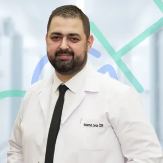
Assoc. Prof. Dr. Muhammet Derda Özer, FICO
Operating Surgeon
Specialises in ocular oncology, vitreoretinal diseases, cataract, refractive, corneal transplantation, glaucoma and pediatric ophthalmic surgery — more than 30,000 ophthalmic operations across a 14-year clinical career.
View full profile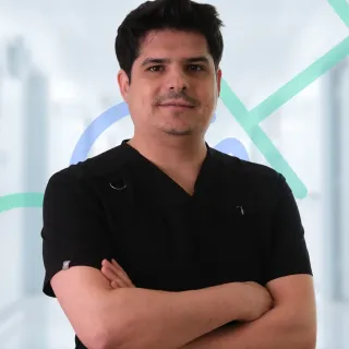
Op. Dr. Önder Aslan
Operating Surgeon
Board-certified ophthalmologist whose surgical practice spans anterior-segment procedures with oculoplastic and retinal sub-specialties — cataract, refractive, eyelid surgery and pediatric eye examinations including retinopathy of prematurity.
View full profile
Op. Dr. Muhammed Talha Sadık
Operating Surgeon
Board-certified ophthalmologist working across vitreoretinal disease, refractive, cataract and glaucoma surgery, with an active anterior- and posterior-segment surgical caseload.
View full profileAre you a candidate for ptosis surgery?
You may be a good candidate if
- You have confirmed blepharoptosis — your upper eyelid consistently covers the pupil or significantly reduces your visual field, documented by marginal reflex distance measurement (MRD-1 ≤2 mm is the standard threshold for functional ptosis).
- You have aponeurotic ptosis — age-related thinning or disinsertion of the levator aponeurosis, typically with normal or good levator function (≥8 mm LF) and a high or absent lid crease.
- You have congenital ptosis from birth with measurable levator function and no amblyopia currently managed under a paediatric ophthalmologist.
- You have neurogenic ptosis from Horner syndrome (confirmed with cocaine/apraclonidine test) or incomplete third nerve palsy with stable neurological workup.
- You have myogenic ptosis from chronic progressive external ophthalmoplegia (CPEO) or myasthenia gravis in remission, with neuro-ophthalmic clearance.
- You are seeking combined ptosis repair and upper blepharoplasty where both a functional levator deficit and significant dermatochalasis coexist.
Surgery should be deferred or reconsidered if
- Active or incompletely worked-up neurogenic ptosis — ptosis from third nerve palsy, myasthenia gravis, or unexplained sudden-onset ptosis requires neuro-ophthalmic investigation and a stable period before elective surgery.
- Absent or severely reduced Bell's phenomenon — if the eye does not rotate upward adequately on lid closure, aggressive ptosis correction creates a high risk of corneal exposure and ocular surface damage; a more conservative approach is necessary.
- Severe dry eye syndrome not yet managed — ptosis repair reduces lid closure range and will worsen dry eye; ocular surface must be treated before surgery.
- Paediatric ptosis with active amblyopia currently under treatment — the amblyopia plan should be coordinated with a paediatric ophthalmologist before surgical timing is decided.
- Patients seeking ptosis repair purely to achieve a cosmetic appearance change without a measured functional levator deficit — the correct procedure for eyelid aesthetics without ptosis is upper blepharoplasty, not ptosis repair.
Disclaimer. This page is consistent with American Academy of Ophthalmology (AAO) Oculoplastic Section clinical guidelines, American Society of Ophthalmic Plastic and Reconstructive Surgery (ASOPRS) published position statements, and Beard's ptosis classification. It is educational and not a clinical recommendation. Ptosis surgery requires formal levator function measurement and etiology evaluation by a qualified surgeon before any technique is selected — photographs and symptom descriptions alone are insufficient. The Eyeglow consultation is provided at no charge and without obligation.
Realistic outcomes — the risks that matter
Ptosis surgery has a good safety record in skilled hands, but its complications are different from standard eyelid procedures. Our oculoplastic surgical team explains these in full during the pre-operative consultation:
Asymmetry requiring revision
Mild height asymmetry between the two upper eyelids occurs in 10 to 20 percent of bilateral cases as swelling resolves unequally. Most asymmetry settles within 4 to 8 weeks without intervention. If a clinically significant asymmetry remains at three months, a minor in-office adjustment is performed. Major revision requiring full re-operation occurs in under 5 percent of carefully selected cases.
Lagophthalmos (incomplete lid closure)
Incomplete closure during sleep or blink is an expected short-term consequence of ptosis repair — the lid is being advanced or tightened. Nocturnal lubricating drops or ointment are prescribed routinely for the first month. Clinically significant lagophthalmos beyond 2 mm at one month that does not resolve with lubrication requires adjustment. Bell's phenomenon assessment before surgery identifies higher-risk corneas.
Under-correction (ptosis recurrence)
Inadequate correction occurs in 5 to 15 percent of cases and is most common in congenital ptosis with poor levator function or in advanced aponeurotic ptosis with very thin tissue. Intraoperative adjustability under local anaesthesia and careful pre-operative grading reduce but do not eliminate this risk. Secondary surgery to advance the levator further or convert to a frontalis sling is straightforward.
Over-correction (lid retraction)
Over-elevation of the lid, causing a startled or wide-eye appearance, occurs in 3 to 8 percent of cases. Early intervention (within 2 weeks while sutures are accessible) can release the levator advancement. If caught late, a small lid-lowering procedure is required. This risk is minimised by intraoperative height assessment under local anaesthesia, which is standard at Eyeglow for levator advancement cases.